Why Do I Need an MRI? Is a Standard X-Ray Not Enough?
Why Do I Need an MRI? Is a Standard X-Ray Not Enough?
When patients come in with radiating back pain, one of the most common questions I hear is, "Doctor, I just had an X-ray at the clinic. Why do I need an MRI too? Isn't it redundant?"
I completely understand this concern. An MRI takes time and involves additional costs. Today, I want to humbly share my perspective and explain the difference between these two imaging tools in the simplest terms possible.
1. The X-Ray: Looking at the Building's Framework
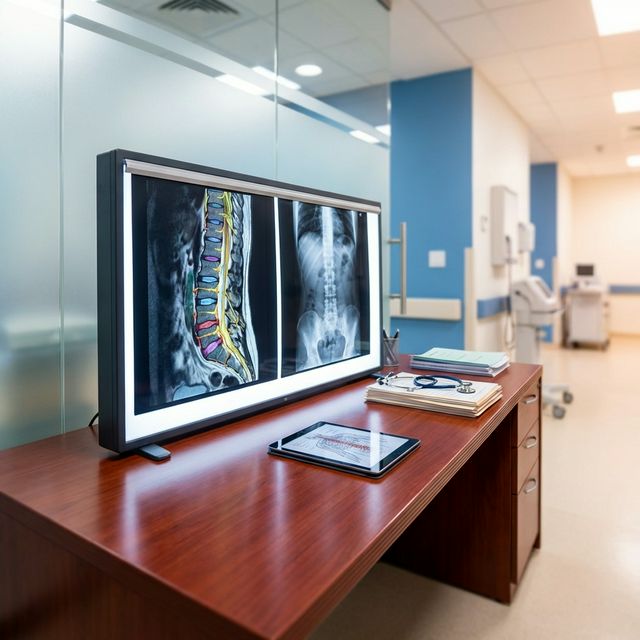
Think of an X-ray as looking at the structural framework of a building. X-rays are excellent at capturing high-density materials, which in our body means bones.
- What an X-ray shows: Are there any fractures? Is the spine aligned properly, or is it slipping? Are there large bone spurs?
- What an X-ray cannot show: Discs, nerves, muscles, and ligaments. The radiation simply passes right through these soft tissues, rendering them invisible on the film.
2. The MRI: Seeing the Wiring Inside the Walls
An MRI (Magnetic Resonance Imaging) uses strong magnetic fields and radio waves to create highly detailed images. It is like being able to look through the walls to see the delicate wiring and plumbing of the building.
- What an MRI shows: Exactly how much a spinal disc has herniated, which specific nerve root is being pinched, the water content of your discs (indicating their health), and if there are any hidden infections or tumors.
Why Endoscopic Surgery Absolutely Requires an MRI
Endoscopic Spine Surgery is performed through an incision the size of a fingernail (about 8 millimeters). Because the incision is so small, there is no room for the surgeon to "look around" to find the problem once inside.
Therefore, we must know the precise location of the nerve compression down to the millimeter before the surgery even begins. The MRI acts as our ultra-precise GPS, allowing us to plan the exact trajectory for the camera tube. This ensures the procedure is highly targeted, exceptionally safe, and causes minimal disruption to your body.
However, as all patients' conditions are varied and the decision-making process should be individualized, the feasibility of doing spinal endoscopy might not be the same for each patient. Consultation with a spine specialist is strongly recommended.
References
- Jarvik, J. G., & Deyo, R. A. (2002). "Diagnostic evaluation of low back pain with emphasis on imaging." Annals of Internal Medicine. [Link]
- Wassenaar, M., et al. (2012). "Magnetic resonance imaging in the diagnosis of radiculopathy: a systematic review." European Spine Journal. [Link]
- Boden, S. D., et al. (1990). "Abnormal magnetic-resonance scans of the lumbar spine in asymptomatic subjects. A prospective investigation." The Journal of Bone and Joint Surgery (American Volume). [Link]

Questions about your symptoms?
Chat with us on LINE for more information
@bright.endospine
Chat on LINE