Endoscopic Surgery for a Cervical Disc Herniation & Foraminal Stenosis (Posterior Cervical Foraminotomy/Discectomy) — a Motion-Preserving Approach
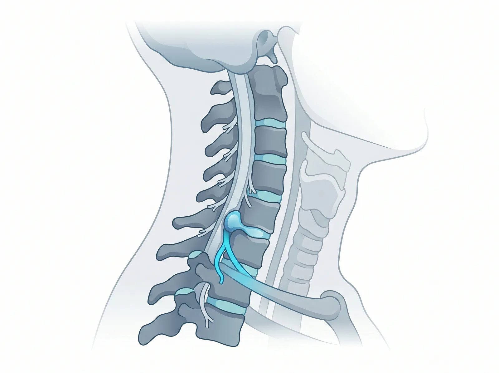
Neck pain that radiates down the arm, along with numbness or weakness in the arm or hand, is often caused by compression of a spinal nerve in the neck. This happens in two main patterns: a cervical disc herniation pressing on a nerve, and narrowing of the bony opening the nerve passes through (cervical foraminal stenosis). When medication, physical therapy, and activity changes have been tried fully and symptoms still interfere with daily life, surgery may be considered. For selected patients, one of the less invasive options available today is posterior endoscopic cervical foraminotomy/discectomy (PECF/PECD) — performed through a small incision at the back of the neck. The defining concept of this approach is that it is motion-preserving: because it does not fuse the spinal segment, the neck joint at that level keeps moving naturally. It is important to be clear, though, that this approach has specific, narrower indications than the standard anterior operation — anterior cervical discectomy and fusion (ACDF). In this article I want to explain, in plain language, what this operation is, who is and isn't a good candidate, how it is done, and how to care for yourself afterward.
What is posterior endoscopic cervical surgery — and why is it "motion-preserving"?
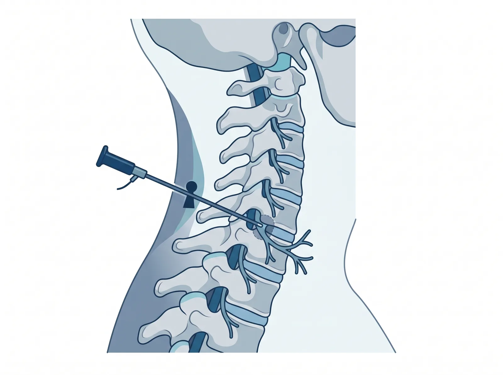
The principle is to pass a high-definition endoscope through a small incision at the back of the neck, reach the bony opening the nerve travels through, and then open up the nerve's channel (foraminotomy) and/or remove just the portion of disc that is compressing the nerve (discectomy) — relieving the pressure while disturbing the surrounding muscle and structures as little as possible. A continuous saline irrigation system keeps the view of the nerve clear throughout.
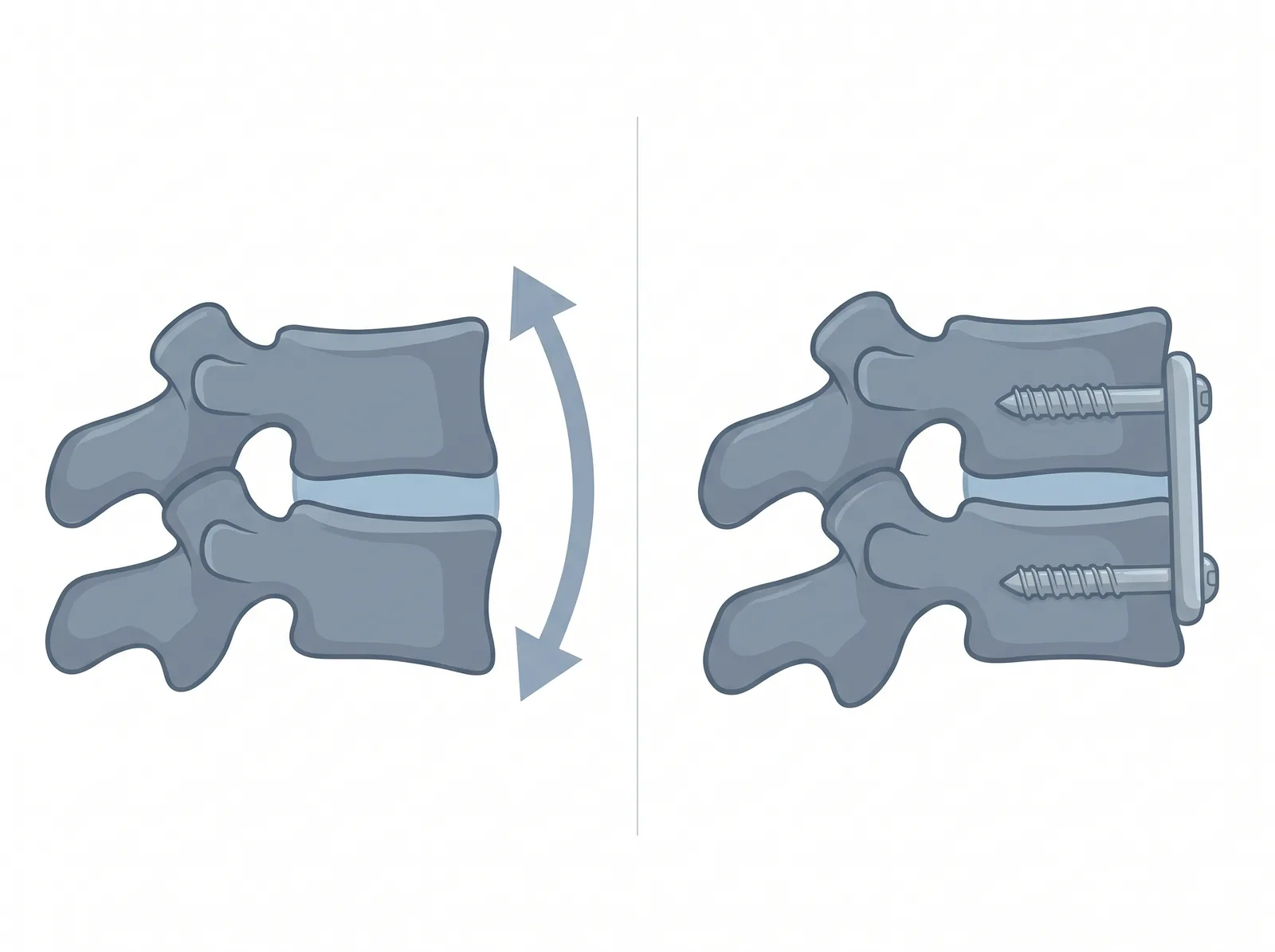
What sets this approach apart is that it does not remove the whole disc and does not fuse the segment, so the neck joint at that level still moves naturally — unlike ACDF, which joins two vertebrae together. This motion preservation is a property inherent to the technique (not a guaranteed result), and it is a key reason some patients are suited to it.
Why motion preservation matters — and the trade-off of fusion (ACDF)
First, to be clear: anterior cervical discectomy and fusion (ACDF) is a good, standard operation and is genuinely necessary for many patients — particularly when there is central cord compression, symptomatic cord compression (myelopathy), cervical instability, or multilevel disease. In those situations, fusion is the more appropriate and safer choice. This article is not saying one approach is "better" overall — each has its own indications.
That said, fusion is not always the end of the story. When one cervical segment is fused and no longer moves, more of the movement load is shifted onto the neighbouring (adjacent) levels. Over time, that can accelerate degeneration of the adjacent level — a condition called adjacent segment disease (ASD). In some patients this can bring symptoms back years later, and some may eventually need another (revision) fusion. Long-term studies following patients after ACDF have shown this trend clearly (see the Hilibrand 1999 reference).
This is exactly why, in patients whose pathology allows it (for example a lateral / foraminal focal compression), having a motion-preserving option such as the posterior endoscopic approach — which does not fuse the segment — is valuable: it lets that level keep moving naturally and avoids a fusion. To be clear again, this approach is not a substitute for ACDF in every case, and suitability has to be assessed individually.
For the big picture of endoscopic spine surgery, see the overview; and to understand a cervical herniated disc itself, see
Who it is for — and who it is NOT for (the honest version — the most important section)
This approach is not right for every patient or every pathology. Its indications are narrow and depend heavily on how the nerve is being compressed. Choosing the right patient matters just as much as the surgical technique itself.
Often suitable when:
- A lateral / foraminal soft cervical disc herniation is compressing a single nerve, causing clear radiating arm pain, with a location that matches the MRI.
- Cervical foraminal stenosis is pinching a nerve at a specific spot, causing arm symptoms.
- Appropriate non-surgical care has been tried for a reasonable period and symptoms still disrupt daily life.
Not the right choice — and where anterior ACDF (fusion) remains the standard — in situations such as:
- Central canal stenosis that primarily compresses the spinal cord.
- Myelopathy (symptomatic cord compression) — e.g. clumsy hands, unsteady walking, worsening balance.
- Cervical instability that requires fusion.
- Multilevel disease or significant cervical deformity.
In this second group, anterior cervical discectomy and fusion (ACDF) remains the standard operation, with safe and appropriate results — the posterior endoscopic approach is not a substitute for it in every case. On indications — when endoscopy is not the right call — see
How it is done (the approach)
Details are individualized, but the general flow is:
- Anesthesia — selected for each patient together with the anesthesiologist.
- A small incision at the back of the neck — the muscle fibers are gently dilated (not stripped over a wide area).
- Guiding the endoscope to the compression — using MRI planning and intra-operative X-ray for positioning.
- Opening the nerve's channel and/or removing the compressing disc fragment — under high-definition magnification and continuous irrigation, relieving pressure on the nerve, without fusing the segment.
- Closure — because the incision is small, only minimal closure is usually needed.
Endoscopic surgery relies on a precise MRI to plan the trajectory and to confirm whether the compression suits this approach — see
Recovery — what to realistically expect
- Most patients are able to get up and walk relatively soon after surgery, and typically have a short hospital stay (depending on the individual and the surgeon's assessment).
- Radiating arm pain often improves gradually; long-standing numbness or weakness may recover at different rates in different people.
- Activity should be increased step by step, following post-operative guidance closely — especially neck movement in the early period.
For post-op care, returning to driving, and (for international patients) how long to rest before flying home, see
Risks & honest limits (educational, not alarmist)
Every operation carries risk, even with a small incision. Risks and limits to be aware of include:
- Recurrence of symptoms: surgery fixes the mechanical compression at that spot but does not stop the natural degeneration of the spine, so symptoms can return — this is not a "permanent cure."
- Dural tear / CSF leak: uncommon, but possible.
- Injury to a nerve or the spinal cord: reported as very uncommon in the literature, but it is a risk that must always be considered in surgery around the neck.
- Incomplete decompression, or finding the pathology is not suited to the endoscopic approach — which in some cases means converting to a different operation, such as anterior ACDF.
FAQ
How is posterior endoscopic cervical surgery different from fusion (ACDF)?
The main difference is that the posterior endoscopic approach aims to relieve compression at a specific spot without fusing the segment, so the neck joint at that level keeps moving — whereas ACDF is an anterior approach that joins the vertebrae together. Both have their own indications: the endoscopic approach suits certain focal compressions, while ACDF remains the standard for central cord compression, instability, or multilevel disease.
Can every cervical disc herniation be treated with the posterior endoscopic approach?
No. It suits certain lateral / foraminal compressions more than others. If the compression is central or there is cord compression (myelopathy), a different operation is usually needed. Individualized assessment from the MRI is essential.
Indications for EndoscopyWhat does "motion-preserving" mean?
Because the segment is not fused, the neck joint at that level still moves naturally — a property inherent to the technique (not a guarantee of outcome). Whether it is appropriate depends on how each person's compression looks.
I have hand numbness, unsteady walking, and worse balance — is this approach right for me?
Those symptoms can be signs of cord compression (myelopathy), which is usually not an indication for the posterior endoscopic approach, and should be assessed by a spine specialist promptly.
(Every case is different and decisions should be individualized. Consultation with a spine specialist is strongly recommended to assess your actual symptoms.)
References
- Ruetten, S., Komp, M., Merk, H., & Godolias, G. (2008). "Full-endoscopic cervical posterior foraminotomy for the operation of lateral disc herniations using 5.9-mm endoscopes: a prospective, randomized, controlled study." Spine (Phila Pa 1976). [Link]
- Kim, C. H., Kim, K. T., Chung, C. K., Park, S. B., Yang, S. H., Kim, S. M., & Sung, J. K. (2015). "Minimally invasive cervical foraminotomy and diskectomy for laterally located soft disk herniation." European Spine Journal. [Link]
- Alomar, S. A., Maghrabi, Y., Baeesa, S. S., & Alves, Ó. L. (2022). "Outcome of Anterior and Posterior Endoscopic Procedures for Cervical Radiculopathy Due to Degenerative Disk Disease: A Systematic Review and Meta-Analysis." Global Spine Journal. [Link]
- Hilibrand, A. S., Carlson, G. D., Palumbo, M. A., Jones, P. K., & Bohlman, H. H. (1999). "Radiculopathy and myelopathy at segments adjacent to the site of a previous anterior cervical arthrodesis." J Bone Joint Surg Am. [Link]
- Kwok, W. C. H., Wong, C. Y. Y., Law, J. H. W., Tsang, V. W. T., Tong, L. W. L., Samartzis, D., An, H. S., & Wong, A. Y. L. (2022). "Risk Factors for Adjacent Segment Disease Following Anterior Cervical Discectomy and Fusion with Plate Fixation: A Systematic Review and Meta-Analysis." J Bone Joint Surg Am. [Link]

Questions about your symptoms?
Chat with us on LINE for more information
@bright.endospine
Chat on LINE